Finding accredited CPD
This course focuses on the approach to pigmented skin lesions relating to skin cancer medicine. Participants will acquire the knowledge required to safely and confidently diagnose and treat commonly encountered skin lesions. It is the ideal starting point to build core knowledge in skin cancer management and acquire vital diagnostic skills and basic management techniques to provide effective care to patients.
Unit one discusses common benign lesions including freckles, solar lentigo, seborrheic keratosis, haemangioma, dermatofibroma and blue nevi. Normal and dermoscopic clinical images are provided to demonstrate patterns and assists with analysis. Benign lesions can mimic skin cancers and careful examination is required to decide management steps. The signs of common benign lesions that may be skin cancer are discussed in detail. A medico-legal case is included for a patient where a melanoma was masquerading as a benign lesion.
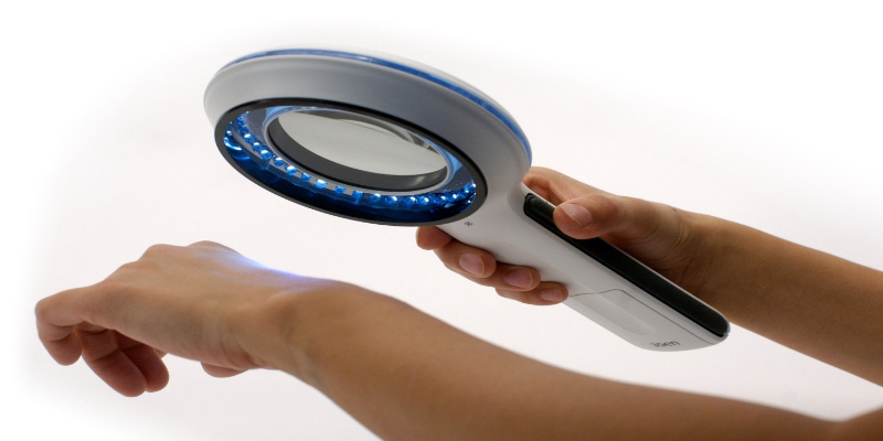
Unit two begins with the description of dermoscopy including examples of dermoscopic devices used for this technique. The 3-point checklist consists of asymmetry in colour or structures, atypical network and blue-white structures (white scar-like depigmentation or blue pepper-like, globular or structure-less areas), showing various clinical dermoscopic images of lesions.
This unit covers dysplastic nevi and melanoma. A table is shown of the relationship between nevus, dysplastic nevus and melanoma and includes a table of relative risk factors for melanoma. Several types of melanomas and treatment options are listed and supported by clinical images. The unit concludes with key points for detecting benign or suspicious lesions.
Cost: $195
Suitable for: All degree qualified medical practitioners.
Study mode: 100% online
Disclaimer: Please note, once you click 'Register now' you will be leaving the AMA’s CPD Home website and entering a third-party education provider’s website. If you choose to register for this learning, you will need to provide some of your personal information directly to the third-party education provider. If you have any queries about how third-party education providers use, disclose or store your personal information you should consult their privacy policy.
Upon completion, your CPD activity record may take up to 4 weeks to be reflected on your CPD Home Dashboard.
You have to log in to see the content of this module.
Provided by

Accredited by