Finding accredited CPD
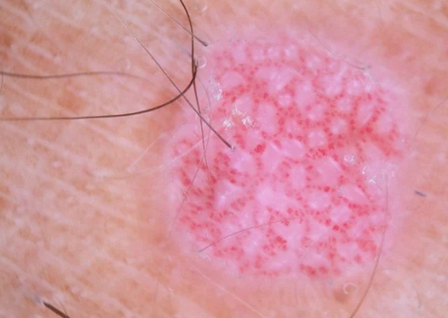
The Dermoscopy for Pink Lesions course equips healthcare professionals with specialised knowledge and practical techniques to diagnose and manage non-pigmented skin tumours effectively. The single-unit program delivers a systematic approach to analysing vascular patterns and clinical presentations of pink lesions.
The course provides an in-depth exploration of the dermoscopic examination of pink lesions, focusing on melanocytic and non-melanocytic tumours. Participants will learn a structured methodology to improve diagnostic confidence.
Key topics include:
- General Principles of Examining Pink Lesions:
- Differentiating inflammatory from tumoral lesions through clinical examination
- The importance of vessel morphology, arrangement, and associated clues
- Four-Step Approach to Pink Lesion Analysis:
- Clinical evaluation to distinguish between tumour types
- Assessment of vascular patterns, including comma, dotted, hairpin, and arborizing vessels
- Arrangement patterns: regular, radial, clustered, or polymorphic
- Dermoscopic patterns for specific tumours
- Dermoscopy Patterns for Specific Tumours:
- Melanocytic tumours (e.g., melanoma, Spitz nevi) with vascular polymorphism and pigmentation clues.
- Non-melanocytic tumours, including basal cell carcinoma, squamous cell carcinoma, sebaceous hyperplasia, and Bowen's disease
- Management Rules for Pink Lesions:
- Recognising high-risk features such as residual pigmentation, ulceration, and polymorphic vessels
- Guidelines for excision to avoid misdiagnosing malignant lesions
- Role of polarised dermoscopy in detecting subtle malignancy indicators like chrysalis structures.
Through interactive case discussions and dermoscopic imagery, participants will gain practical skills for identifying benign and malignant lesions confidently.
This course is essential for practitioners looking to enhance their proficiency in dermoscopy, particularly for challenging pink lesions, ensuring better patient outcomes through accurate diagnosis and effective management.
Cost: $195
Suitable for: All degree qualified medical practitioners.
Study mode: 100% online
Disclaimer: Please note, once you click 'Register now' you will be leaving the AMA’s CPD Home website and entering a third-party education provider’s website. If you choose to register for this learning, you will need to provide some of your personal information directly to the third-party education provider. If you have any queries about how third-party education providers use, disclose or store your personal information you should consult their privacy policy.
Upon completion, your CPD activity record may take up to 4 weeks to be reflected on your CPD Home Dashboard.
You have to log in to see the content of this module.
Provided by

Accredited by